Pericardial involvement in neoplastic disease: case report

Neda Daukšaitė1, Gintarė Valterytė1, Laura Marcinkienė3, Vytautas Zabiela2, Rita Kupčinskaitė-Noreikienė3, Diana Žaliaduonytė2

1Medical Academy, Lithuanian University of Health Sciences, Kaunas, Lithuania.

2Department of Cardiology, Medical Academy, Lithuanian University of Health Sciences,

Kaunas, Lithuania.

3Department of Hematology and Oncology, Medical Academy, Lithuanian University of  Health Sciences, Kaunas, Lithuania.


Abstract

1.1 Background

Primary tumors of the pericardium are rare. Secondary or metastatic pericardial disease is much more common. Symptomatic pericarditis may be the first clinical manifestation of malignancy. The diagnosis is usually an incidental finding during imaging processing. Pericardiocentesis and percutaneous drainage of pericardial effusion (PE) are indicated in PE if neoplastic etiology is suspected. In this report, we describe the case of a patient presenting with nonspecific symptoms of PE and neoplastic disease.

1.2 Case presentation

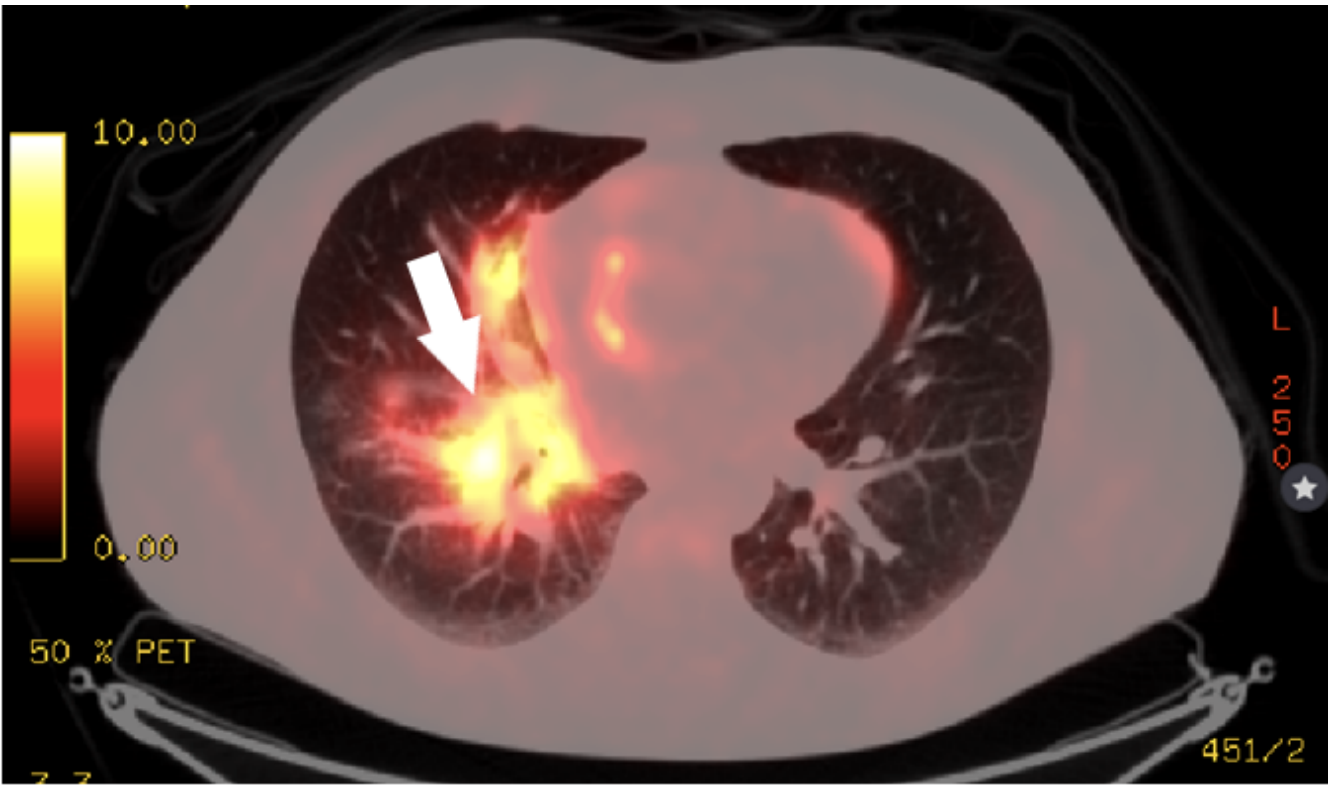
A 47 – year old man was admitted to the hospital with 2 years history of intermittent low – grade fever, non – productive cough, malaise, and dyspnea. Computed tomography (CT) scan was done allegedly to find the pulmonary embolism, but the CT scan disclosed multiple enlarged lymph nodes in mediastinal, intraperitoneal areas, and pericardial effusion. Pericardial effusion and neoplastic disease were diagnosed by lymph node biopsy and positron emission tomography/computed tomography (PET/CT), respectively. Treatment started with Alectinib and radiation therapy. However, after 10 months the progression of the disease was observed, so treatment changed to second-line drug Lorlatinib. In order to prevent pathological bone fractures, treatment changed to Denosumab. It resulted in the complete remission of PE. In 1 year and 6 months after diagnosis of the lung cancer was confirmed regarding the successful treatment, the patient is fully active, back to social life with no signs of dyspnea.

1.3 Conclusions

The diversity of clinical manifestation (such as low – grade fever, non – productive cough, malaise, dyspnea, and PE) in such a potentially severe disease should alert the physician to prompt diagnosis and treatment of malignant process.

1.4 Keywords:  Pericarditis; Effusion; Neoplastic disease; Case; Malignancy.


Full article

Journal of Medical Sciences. April 30, 2020 - Volume 8 | Issue 15. Electronic-ISSN: 2345-0592
257
Medical Sciences 2020 Vol. 8 (15), p. 257-268
Pericardial involvement in neoplastic disease: case report
Neda Daukšaitė
1
, Gintarė Valterytė
1
, Laura Marcinkienė
3
, Vytautas Zabiela
2
, Rita Kupčinskaitė-
Noreikienė
3
, Diana Žaliaduonytė
2
1
Medical Academy, Lithuanian University of Health Sciences, Kaunas, Lithuania.
2
Department of Cardiology, Medical Academy, Lithuanian University of Health Sciences,
Kaunas, Lithuania.
3
Department of Hematology and Oncology, Medical Academy, Lithuanian University of Health
Sciences, Kaunas, Lithuania.
Abstract
1.1 Background
Primary tumors of the pericardium are rare. Secondary or metastatic pericardial disease is much more common. Symptomatic
pericarditis may be the first clinical manifestation of malignancy. The diagnosis is usually an incidental finding during imaging
processing. Pericardiocentesis and percutaneous drainage of pericardial effusion (PE) are indicated in PE if neoplastic etiology is
suspected. In this report, we describe the case of a patient presenting with nonspecific symptoms of PE and neoplastic disease.
1.2 Case presentation
A 47 - year old man was admitted to the hospital with 2 years history of intermittent low - grade fever, non - productive cough,
malaise, and dyspnea. Computed tomography (CT) scan was done allegedly to find the pulmonary embolism, but the CT scan
disclosed multiple enlarged lymph nodes in mediastinal, intraperitoneal areas, and pericardial effusion. Pericardial effusion and
neoplastic disease were diagnosed by lymph node biopsy and positron emission tomography/computed tomography (PET/CT),
respectively. Treatment started with Alectinib and radiation therapy. However, after 10 months the progression of the disease was
observed, so treatment changed to second-line drug Lorlatinib. In order to prevent pathological bone fractures, treatment changed
to Denosumab. It resulted in the complete remission of PE. In 1 year and 6 months after diagnosis of the lung cancer was
confirmed regarding the successful treatment, the patient is fully active, back to social life with no signs of dyspnea.
1.3 Conclusions
The diversity of clinical manifestation (such as low - grade fever, non - productive cough, malaise, dyspnea, and PE) in such a
potentially severe disease should alert the physician to prompt diagnosis and treatment of malignant process.
1.4 Keywords: Pericarditis; Effusion; Neoplastic disease; Case; Malignancy.
Journal of Medical Sciences. April 30, 2020 - Volume 8 | Issue 15. Electronic-ISSN: 2345-0592
258
2. Introduction
Malignant involvement of the pericardium is detected in 1
to 20 percent of cancer cases in autopsy studies (1).
However, primary tumors of the pericardium are rare.
Secondary or metastatic pericardial disease is much more
common and arises in most cases from lung, breast tumors,
melanoma, or hematologic malignancies (2). Symptomatic
pericarditis, pericardial effusion, and sometimes even
cardiac tamponade may be the first clinical manifestation of
malignancy. The clinical suspicions of neoplastic
involvement of the pericardium are not straightforward and
symptoms such as dyspnea, tachycardia, and chest pain are
not specific and may be caused by anemia or pleural
effusion in cancer patients. The diagnosis is usually an
incidental finding during imaging processing. The thoracic
X-ray can be valuable and show the presence of an enlarged
cardiac silhouette with clear lungs. These findings should
raise the suspicion of PE. However, they provide
nonspecific information, and an echocardiogram should be
done. The echocardiogram is the most available and reliable
technique in order to verify the presence and the amount of
a PE (3). There are 10 echocardiographic findings described
in cardiac tamponade due to neoplastic PE. The most
common ones are signs of ventricular interdependence:
abnormal inspiratory increase of right ventricle (RV)
dimensions and abnormal inspiratory decrease of left
ventricle (LV) dimensions; right atrium (RA) systolic
collapse or RV early diastolic collapse; diastolic reversal of
hepatic vein flow on expiration; a plethora of the inferior
vena cava and swinging motion of the heart in the
pericardial sac (3, 4). A CT is an equally useful imaging
tool and may identify PE in patients that first present with
dyspnea and characterize the pericardial fluid in cases of
unknown etiology (5). Cardiac catheterization may be
considered when there is a discrepancy between the clinical
and non - invasive imaging, and in patients with complex
hemodynamic disorders. It reveals elevated RA pressure,
with a prominent x descent and a diminished or absent y
descent on the RA pressure curve (6). Pericardiocentesis
and percutaneous drainage of PE is life - saving in
tamponade and is indicated in symptomatic moderate to
large PE refractory to medical therapy or if bacterial or
neoplastic etiology is suspected (7, 8). If the PE is
hemorrhagic, malignancy should always be considered.
3. Case: A 47 - year old man was admitted to the Hospital
of Lithuanian University of Health Sciences, Kaunas clinics
with 2 years history of intermittent low - grade fever, non -
productive cough, malaise, and dyspnea. Subsequently, the
patient’s condition began to worsen so he was brought to
the local emergency room for evaluation. Physical
Journal of Medical Sciences. April 30, 2020 - Volume 8 | Issue 15. Electronic-ISSN: 2345-0592
259
examination revealed subfebrile status, normal heart rate,
elevated blood pressure. Cardiac auscultation revealed a
regular, dull heartbeat without the pericardial friction rub
with a rate of 75 bpm. CT scan was done allegedly to find
the pulmonary embolism. CT scan disclosed multiple
enlarged lymph nodes in mediastinal, intraperitoneal areas,
and PE (PE up to 26 mm). The pericardiocentesis was
performed and bloody fluid was drained from the
pericardial space. However, the analysis of pericardial fluid
was not informative enough. The patient was subsequently
transferred to the cardiology department of our hospital for
further evaluation.
Laboratory tests: blood test showed normocytic
normochromic anemia: red blood cells 3.85 x 10
12
/l (as
normal value is 4.4 - 5.6 x 10
12
/l), hemoglobin 116 g/l
(normal value is 135 - 169 g/l), hematocrit 34.10% (normal
value is 40% - 49%), MCV 88.60 fl (normal value is 82 -
96 fl), MCH 30.10 pg (normal value is 27 - 32.3 pg),
neutrophils 7.10 x 10
9
/l (normal value is 1.8 - 7.4 x 10
9
/l),
platelets 329 x 10
9
/l (normal value is 166 308 x 10
9
/l).
The leukocytosis was observed as white blood cell count
was 9.63 x 10
9
/l (normal value is 3.9 - 8.8 x 10
9
/l). C-
reactive protein (CRP) was elevated up to 70.14 mg/l (when
normal value is 0 - 7.5 mg/l). All the blood clotting tests
were normal.
An electrocardiogram (ECG) showed reduced voltage, heart
rate 75 bpm.
Echocardiography revealed the overall separation of
pericardial visceral and parietal layers in diastole
approximately from 9 to 11 mm depending on location (as
shown in Figure 1.). The visceral pericardial tissue of the
right ventricle was thickened, dense, and had even surfaces.
CT of the abdomen and chest was repeated twice and
disclosed multiple enlarged paraaortic lymph nodes up to
2.1 cm in diameter (Figure 2.). The patient was consulted by
a hematologist, oncologist, abdomen surgeon, and the
resection of pathological lymph node was performed as
there were no other pathological findings in the examination
results that could suggest any oncological diagnosis. The
morphologic findings on histopathology combined with the
results of immunohistochemistry confirmed the presence of
metastatic involvement of a prepericardial lymph node by
poorly differentiated adenocarcinoma (the structure of the
lymph node was disrupted and infiltrated with large-scale
tumor cells that had enlarged nucleus and nucleolus,
dispersive chromatin which stained positive for CK18,
CDX2. Tumor cells also showed focal positivity for CK7.
The tumor cells were negative for CD5 and p63 (Figure 5.)).
After the metastasis of adenocarcinoma was found, it was
attempted to discover the primary tumor. For that reason,
esophagogastroduodenoscopy was performed and no
Journal of Medical Sciences. April 30, 2020 - Volume 8 | Issue 15. Electronic-ISSN: 2345-0592
260
pathological changes except for duodenal ulcer were found.
Afterward, fibrocolonoscopy was done and has revealed II°
hemorrhoids without any signs of oncological process. The
consultation of urologist was given as well as the patient
had episodical pain in his testis at the moments of febrile
temperature. No pathological changes in the external genital
and testis area have been found. Tumor markers, such as Ca
19 - 9 (tumor marker in gastrointestinal cancers); CEA
(carcinoembryonic antigen); PSA (prostate-specific
antigen); AFP (alpha-fetoprotein); hCG (human chorionic
gonadotropin), were tested and were found normal. For the
revision of the diagnosis and finding the primary tumor, a
magnetic resonance imaging (MRI) scan of the abdomen
and pelvis was carried out, in which the pathological lymph
nodes were observed: in the retroperitoneum largest up to
3.7 x 3.0 cm, subdiaphragmatic - 1.2 x 1.0 cm. Also, a
thickened stomach wall up to 1.4 cm and metastasis in the
sacrum were found. The performed MRI scan did not help
us found the primary tumor. For the identification of the
primary tumor, in the case of non-informative radiological
tests (CT, MRI), a PET/CT was performed. It identified a
highly metabolically active tumor, spread to the right lung,
liver, bone tissue, neck, mediastinal, and abdominal lymph
nodes, most likely as a primary lung tumor (Figure 3,4.). A
further examination of the histological material determined
anaplastic lymphoma kinase (ALK) translocalization 100%
of cells (+ + +).
At the Cardiology department the patient was treated with
antipyretic drugs, ampicillin - sulbactam for suspected
bacterial pericarditis for a short period. With a complete
resolution of cough, fever and major improvement in CRP
level, antibiotics and antipyretic drugs were stopped. Also, a
treatment of heart failure with metoprolol and torsemide
was prescribed. Omeprazole was prescribed for duodenal
ulcer treatment. Later the patient was transferred to the
Oncology department where the case was discussed at the
Multidisciplinary Oncology board. The first-line treatment
of alectinib p/os was prescribed because of the case of
ALK-positive translocation. At the time of treatment, a
regular thoracic and abdominal CT was performed every 3
months, where a positive dynamic was observed. The
patient was regularly consulted by a cardiologist and the
accumulation of fluid in the pericardium was not observed.
Alectinib treatment was continued for 10 months. However,
the control of the thoracic, abdominal, and pelvic
examinations (after 10 months of treatment with alectinib)
showed the progression of the disease, enlarged mediastinal,
and abdominal lymph nodes. Palliative radiation therapy
was applied to the lymph nodes of the mediastinum. As the
progression of lung adenocarcinoma with alectinib as the
primary treatment was observed, a second-line drug of
Journal of Medical Sciences. April 30, 2020 - Volume 8 | Issue 15. Electronic-ISSN: 2345-0592
261
biological therapy - lorlatinib p/os was chosen. Since
October 2019, treatment with lorlatinib has been initiated,
which continues to date. In order to prevent pathological
bone fractures in the future, treatment with a monoclonal
antibody, denosumab S/C, was additionally used. In 1 year
and 6 months after diagnosis of the lung cancer was
confirmed regarding the successful treatment, the patient is
fully active, back to social life with no signs of dyspnea.
Figure 1. Patient’s transthoracic echocardiocardiography. Apical 4 chamber view revealing
separation of pericardial visceral and parietal layers.
Journal of Medical Sciences. April 30, 2020 - Volume 8 | Issue 15. Electronic-ISSN: 2345-0592
262
.
Figure 2. CT scan of chest and abdomen (transverse view) showing multiple enlarged paraaortic
lymph nodes demonstrated on CT.
Figure 3. PET/CT scan showing the lung tumor and its’ infiltration into mediastinal lymph nodes and
pericardial space.
Journal of Medical Sciences. April 30, 2020 - Volume 8 | Issue 15. Electronic-ISSN: 2345-0592
263
Figure 4. PET/CT scan showing the lung tumor and its’ infiltration into mediastinal lymph nodes.
Figure 5. Immunohistochemistry revealed that lymph node is infiltrated with tumor cells which stained
positive for CK18, CDX2. Tumor cells also showed focal positivity for CK7. The tumor cells were
negative for CD5 and p63.
Journal of Medical Sciences. April 30, 2020 - Volume 8 | Issue 15. Electronic-ISSN: 2345-0592
264
4. Discussion: Our patient did not have classic symptoms
and signs of mild/moderate PE such as pleuritic chest pain,
pericardial friction rubs, and QRS alternans on ECG (9,10).
The main symptoms were dry cough, general weakness, and
intermittent subfebrile temperature. PE was found
accidentally during CT checking for pulmonary
thromboembolism. Therefore, every patient with sudden
dyspnea and significant PE of unspecified etiology should
be examined for possible malignancy (11,12). Pericardial
disease can be the first manifestation of malignancy, more
often when the patient presents with a large PE or
tamponade (11,13). Primary neoplasms of the pericardium
are forty times less common than the metastatic ones (3).
Malignant tumors of the lung, breast, and esophagus as
well as lymphoma, leukemia, and melanoma often
spread to the pericardium directly or through the lymphatic
vessels or bloodstream (13,14). The etiology of PE can be
classified into infectious, autoimmune, iatrogenic,
traumatic, and metabolic neoplastic (15,3). The etiology is
unknown in 40% - 85% of cases (11,3). It is however much
more uncommon for the PE to be the initial presentation of
a hitherto undiagnosed malignancy (16). All of the imaging
tests are diagnostically useful. However, pericardiocentesis
with a cytologic examination of the pericardial fluid should
be performed in patients with a pericardial effusion
whenever there is a reason to suspect malignancy. The
sensitivity of cytology for the diagnosis of a malignant
effusion is between 75% and 92% (17,18). The
pericardiocentesis for our patient was performed and blood-
stained fluid was drained from the pericardial space.
However, fluid analysis in our patient was not informative.
The CT revealed pathologically enlarged lymph nodes at the
paraaortic area and the resection of one of them was
performed. The morphological findings on histopathology
combined with the results of immunohistochemistry
confirmed the presence of metastatic involvement of a
prepericardial lymph node by poorly differentiated
adenocarcinoma. The primary tumor was not detected by
the conventional CT scan, and it is not surprising because
the sensitivity and specificity for lung cancer of this
diagnostic method is only 55% and 81% respectively (19).
PET/CT is more sensitive and specific (80% and 88%,
respectively) diagnostic test, which, in this case, identified a
highly metabolically active tumor, in the right lung spread
in the liver, bone, neck, mediastinal, and abdominal lymph
nodes (19). The usual treatment of large PE and cardiac
tamponade is the drainage of the pericardium, in some cases
intravenous resuscitation in the volume deplete patient may
boost right heart filling pressures, whilst mechanical
ventilation increases intrathoracic pressure thus impeding
Journal of Medical Sciences. April 30, 2020 - Volume 8 | Issue 15. Electronic-ISSN: 2345-0592
265
right-sided filling pressures and can, therefore, be counterproductive.
Metastatic involvement of the pericardium represents a
leading cause of pericardial disease in hospitalized patients
(20). PE and pulmonary embolism associated with
hypercoagulable states are relatively common complications
of malignancy. If malignant PE is sufficiently large to
require drainage, it is a poor prognostic factor, with a
reported median survival of 6.1 months (16). However, new
treatment options seem to offer much better results, as in the
case of our patient. The identification of oncogenic
activation of particular tyrosine kinases in advanced non-
small cell lung cancer (NSCLC) tumors is essentially
important before choosing the treatment tactic because the
tactic can variate, depending on that. The most common
mutations are in the epidermal growth factor receptor
(EGFR), anaplastic lymphoma kinase (ALK) gene, or c -
ROS oncogene-1 (ROS1) gene. Our patient was diagnosed
with ALK translocation. A group of patients with NSCLC
has tumors that contain an inversion in chromosome 2 that
juxtaposes the 5' end of the echinoderm microtubule-
associated protein-like 4 (EML4) gene with the 3' end of the
anaplastic lymphoma kinase (ALK) gene, resulting in the
novel fusion oncogene EML4 - ALK (21). In unselected
NSCLC populations, the ALK rearrangement is a relatively
rare event, on the order of about 4 percent (22). Our patient,
as mainly the other patients with ALK fusion oncogene
positive lung cancer, is male, relatively younger,
nonsmoker, and with histologically confirmed
adenocarcinoma (22). Advanced NSCLC associated with
the ALK fusion oncogene is highly sensitive to ALK
tyrosine kinase inhibitors such as crizotinib, alectinib, and
others. Phase III clinical trials show that anti-ALK therapy
is more effective and is better tolerated compared to
conventional chemotherapy (23,24). Our patient in this case
was treated with alectinib for 10 months, without any
serious adverse events, till the progress of the disease was
diagnosed. As a second - line anti-ALK therapy we have
chosen lorlatinib. It is an effective treatment for patients
with ALK-positive NSCLC who have disease progression
while consuming crizotinib and alectinib (25,26).
5. Conclusions
Intermittent low - grade fever, cough, and malaise might be
not very specific signs of numerous systemic and non -
systemic illnesses. The diagnosis of PE for the patient in
this clinical case was accidental. Low voltage ECG is an
indicative sign of large PE. However, it is important to
perform necessary laboratory or imaging tests, including
pericardial fluid analysis when PE is presented. Whereas in
patients with hemorrhagic pericardial effusions, malignancy
should be always considered.
Journal of Medical Sciences. April 30, 2020 - Volume 8 | Issue 15. Electronic-ISSN: 2345-0592
266
6.References
1. Maisch B, Ristic A, Pankuweit S. Evaluation and
management of pericardial effusion in patients
with neoplastic disease. Prog Cardiovasc Dis.
2010;53(2): 157-63.
2. Mukai K., Shinkai T., Tominaga K., Shimosato Y.
The incidence of secondary tumours of the heart
and pericardium: a 10-year study. Jpn J Clin
Oncol. 1988;18: 195-201.
3. Marwan M.R., William E.K. Neoplastic Pericardial
Effusion. Clin. Cardiol. 2011; 34(10) 593598.
4. Imazio M, Adler Y. Management of pericardial
effusion. Eur Heart J 2013; 34(16), 11861197.
5. Adler Y, Lotan D. Pericardial involvement in
neoplastic disease. In: ESC CardioMed. 3 edn ed.
Oxford, UK: Oxford University Press; 2018:1585-
1587.
6. Adler Y, Ristić AD, Seferović PM, Maisch B,
Kanjuh V. Cardiac tamponade. In: ESC
CardioMed. 3 edn ed. Oxford, UK: Oxford
University Press; 2018:1575-1580.
7. Maisch B, Seferovic PM, Ristic AD, Erbel R,
Rienmüller R, Adler Y, Tomkowski WZ, Thiene
G, Yacoub MH. Guidelines on the diagnosis and
management of pericardial diseases, executive
summary: The Task Force on the diagnosis and
management of pericardial diseases of the
European Society of Cardiology. Eur Heart J 2004;
25(7):587610.
8. Adler Y, Charron P, Imazio M, Badano L, Barón-
Esquivias G, Bogaert J, Brucato A, Gueret P,
Klingel K, Lionis C. et al. 2015 ESC Guidelines
for the diagnosis and management of pericardial
diseases: The Task Force for the Diagnosis and
Management of Pericardial Diseases of the
European Society of Cardiology (ESC)Endorsed
by: The European Association for Cardio-Thoracic
Surgery (EACTS). Eur Heart J 2015; 36(42):2921
2964.
9. Scheinin S., Sosa-Herrera J. Case Report: Cardiac
Tamponade Resembling an Acute Myocardial
Infarction as the Initial Manifestation of Metastatic
Pericardial Adenocarcinoma. Methodist Debakey
Cardiovasc J. 2014 Apr-Jun; 10(2): 124–128.
10. Little WC, Freeman GL. Pericardial disease.
Circulation. 2006; 113:16221632.
Journal of Medical Sciences. April 30, 2020 - Volume 8 | Issue 15. Electronic-ISSN: 2345-0592
267
11. Akhbour S., Khennine BA., Oukerraj L., Zarzur J.,
Cherti M. Pericardial tamponade and coexisting
pulmonary embolism as first manifestation of non-
advanced lung adenocarcinoma. Pan Afr Med J.
2014; 18: 15.
12. Banham-Hall EJ., Bokhari AM. Malignancy with
unknown primary presenting as acute cardiac
tamponade: a case report. Cases Journal 2009,
2:8176.
13. Hamiel U, Yeganeh S, Carrasso S, Soboh S. an
alerting sign: enlarged cardiac silhouette.
Cleve Clin J Med 2015;82:801803.
14. Buzaid AC, Garewal HS, Greenberg BR.
Managing malignant pericardial effusion. West J
Med. 1989;150(2):174179.
15. Atar S, Chiu J, Forrester JS, Siegel RJ. Bloody
pericardial effusion in patients with cardiac
tamponade: is the cause cancerous, tuberculous, or
iatrogenic in the 1990s? Chest. 1999;116(6):1564-
1569.
16. Fong HF., Abdullah O., Gautam S. Case report:
Cough as the sole manifestation of pericardial
effusion. Case Reports 2018;2018: bcr-2017-
222327.
17. Porte HL, Janecki-Delebecq TJ, Finzi L, Métois
DG, Millaire A, Wurtz AJ. Pericardoscopy for
primary management of pericardial effusion in
cancer patients. Eur J Cardiothorac Surg.
1999;16(3):287-291.
18. Saab J, Hoda RS, Narula N, Hoda SA, Geraghty
BE, Nasar A, Alperstein SA, Port JL, Giorgadze T.
Diagnostic yield of cytopathology in evaluating
pericardial effusions: Clinicopathologic analysis of
419 specimens. Cancer Cytopathol.
2017;125(2):128-137.
19. Levy PY, Habib G, Collart F, Lepidi H, Raoult D.
Etiological diagnosis of pericardial effusion.
Future Microbiol. 2006, 1: 229-239.
20. Holt BD. Diseases of the pericardium. In: Fuster V
Alexander RW, O’Rourke RA, Roberts R, King
SB, Nash IS, et al., editors. Hurst’s The Heart. 11th
ed. New York: McGraw-Hill; 2004. p. 1985-95.
21. Shaw AT, Yeap BY, Mino-Kenudson M,
Digumarthy SR, Costa DB, Heist RS, Solomon B,
Stubbs H, Admane S, McDermott U, et al. Clinical
features and outcome of patients with non-small-
cell lung cancer who harbor EML4-ALK. J Clin
Oncol 2009; 27(26):4247-4253.
Journal of Medical Sciences. April 30, 2020 - Volume 8 | Issue 15. Electronic-ISSN: 2345-0592
268
22. Silvestri GA, Gonzalez AV, Jantz MA, et al.
Methods for staging non-small cell lung cancer:
Diagnosis and management of lung cancer, 3rd ed:
American College of Chest Physicians evidence-
based clinical practice guidelines. Chest 2013; 143:
e211S.
23. Shaw AT, Solomon B. Targeting anaplastic
lymphoma kinase in lung cancer. Clin Cancer Res
2011; 17(8):2081-2086.
24. Shaw AT, Kim DW, Nakagawa K, Seto T, Crinó
L, Ahn MJ, De Pas T, Besse B, Solomon BJ,
Blackhall F et al. Crizotinib versus chemotherapy
in advanced ALK-positive lung cancer. N Engl J
Med 2013; 368(25):2385-2394.
25. Novello S, Mazières J, Oh IJ, et al. Alectinib
versus chemotherapy in crizotinib-pretreated
anaplastic lymphoma kinase (ALK)-positive non-
small-cell lung cancer: results from the phase III
ALUR study. Ann Oncol 2018; 29(6):1409-1416.
26. Ojeda W, Martínez-Toro JA. Diagnosis and
management of pericardial effusions. P R Health
Sci J. 2006; 25:255258.
27. Lorlatinib tablets. United States Prescribing
Information. US National Library of Medicine.
http://www.accessdata.fda.gov/drugsatfda_docs/la
bel/2018/210868s000lbl.pdf (Accessed on June 21,
2019).
28. Mirsadraee S
1
, Oswal D, Alizadeh Y, Caulo A, van
Beek E Jr. The 7th lung cancer TNM classification
and staging system: Review of the changes and
implications. World J Radiol. 2012;4(4):128-134.