Pneumonia mimicking invasive pulmonary aspergillosis in patient with lung adenocarcinoma: a case report

Viktor Migunov1

1Vilnius University, Faculty of Medicine

Abstract

Background. Invasive pulmonary aspergillosis is a life-threatening fungal disease and usually affects immunocompromised patients. Clinical symptoms and radiological findings are nonspecific and may be indistinguishable from other pulmonary conditions such as  pneumonia or pulmonary tuberculosis. Invasive pulmonary aspergillosis is a rare condition in patients with solid tumours and is usually  not considered.

Case report. In this case a 58-year-old man was misdiagnosed with pneumonia. After he failed to respond to  antibiotic treatment, pulmonary tuberculosis was suspected. Final diagnosis was invasive pulmonary aspergillosis and lung adenocarcinoma. This case illustrates the challenges of recognizing invasive pulmonary aspergillosis.

Discussion. Diagnosis of invasive pulmonary aspergillosis is challenging and requires a combination of clinical, radiological and microbiological features. Diagnostic methods and  accuracy in recognizing invasive pulmonary aspergillosis can differ and depend on patients clinical features.

Keywords. Aspergillus, invasive pulmonary aspergillosis, tuberculosis, lung adenocarcinoma.

Journal of Medical Sciences. Mar 18, 2022 - Volume 10 | Issue 1. Electronic - ISSN: 2345-0592
150
Medical Sciences 2021 Vol. 10 (1), p. 150-156, https://doi.org/10.53453/ms.2022.03.17
Pneumonia mimicking invasive pulmonary aspergillosis in
patient with lung adenocarcinoma: a case report
Viktor Migunov
1
1
Vilnius University, Faculty of Medicine
Abstract
Background. Invasive pulmonary aspergillosis is a life-threatening fungal disease and usually affects
immunocompromised patients. Clinical symptoms and radiological findings are nonspecific and may be
indistinguishable from other pulmonary conditions such as pneumonia or pulmonary tuberculosis.
Invasive pulmonary aspergillosis is a rare condition in patients with solid tumours and is usually not
considered.
Case report. In this case a 58-year-old man was misdiagnosed with pneumonia. After he failed to respond
to antibiotic treatment, pulmonary tuberculosis was suspected. Final diagnosis was invasive pulmonary
aspergillosis and lung adenocarcinoma. This case illustrates the challenges of recognizing invasive
pulmonary aspergillosis.
Discussion. Diagnosis of invasive pulmonary aspergillosis is challenging and requires a combination of
clinical, radiological and microbiological features. Diagnostic methods and accuracy in recognizing
invasive pulmonary aspergillosis can differ and depend on patients clinical features.
Keywords. Aspergillus, invasive pulmonary aspergillosis, tuberculosis, lung adenocarcinoma.
Journal of Medical Sciences. Mar 18, 2022 - Volume 10 | Issue 1. Electronic - ISSN: 2345-0592
151
Introduction
Invasive aspergillosis is a life-threatening fungal
disease which usually affects
immunocompromised patients. It’s mostly
reported in patients with hematologic
malignancies, stem cell and solid organ
transplant recipients, other risk factors include
prolonged therapy with high-dose
corticosteroids, cytotoxic therapy, advanced
AIDS, chronic granulomatous disease (1-3).
Clinical symptoms of invasive pulmonary
aspergillosis (IPA) and radiological findings are
nonspecific and may be indistinguishable from
pneumonia which can lead to delay in diagnosis
and potentially worse outcomes (4). IPA is often
not considered in patients with solid tumours (5)
and this clinical case illustrates that IPA should
be considered in oncological patients,
particularly those with lung cancer and having
pneumonia symptoms.
Case report
A 58-year-old man went to the primary care
physician with complaints of cough, fever, left
side chest pain and arthralgias lasting for one
week. Patient has about thirty pack-year
smoking history and is without any underlying
diseases. On physical examination the patient
presented with fever 38,5 °C, auscultation of the
chest revealed left-sided coarse crackles in the
upper zone, other vital signs were normal. White
blood cell count (WBC) was 19 ´ 10
9
/L (4 - 10
´ 10
9
/L), C-reactive protein (CRP) 90 mg/l (<
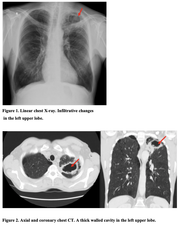
5mg/l). Chest X-ray revealed infiltrative
changes in the left upper lobe pulling left lung
root upward (Fig.1). Patient was diagnosed with
pneumonia and discharged home with prescribed
oral antibiotic treatment (amoxiclav
875/125mg). Ten days later with no clinical
improvement the patient was referred to a
pulmonologist for consultation suspecting a
pulmonary tuberculosis (PTB). On examination
the patient presented with fever (38.5 °C),
fatigue and worsening left side chest pain. WBC
34 ´ 10
9
/L (4 - 10 ´ 10
9
/L), CRP 90mg/l (<
5mg/l). Chest X-ray without essential dynamics.
The patient was suspected of having PTB,
though microscopical, cytological and molecular
(Xpert MTB/RIF) test of sputum smear for TB
was negative. Chest computed tomography
revealed a thick walled cavity (66 ´ 45 mm) in
the left upper lobe and mediastinal
lymphadenopathy (Fig.2). Fibrobronchoscopy
was performed and showed bloody-purulent
secretion in the left upper lobe. Bronchial
aspirate sample was taken for Tuberculosis
mycobacterium exclusion and all tests were
negative. Diagnostic biochemical test revealed a
positive galactomannan (GM) test (index >3.5)
from broncho-alveolar lavage (BAL) also
microbiological culture for Aspergillus
fumigatus was positive. Patient was diagnosed
with invasive pulmonary aspergillosis (IPA) and
treated with Voriconazole 200mg twice a day for
two weeks and then underwent surgical
treatment - left lung upper lobe S1 - S2 resection.
Histological examination of resected tissues was
classified as a poorly differentiated G3 lung
adenocarcinoma pT1b pN1.
Journal of Medical Sciences. Mar 18, 2022 - Volume 10 | Issue 1. Electronic - ISSN: 2345-0592
152
Figure 1. Linear chest X-ray. Infiltrative changes
in the left upper lobe.
Figure 2. Axial and coronary chest CT. A thick walled cavity in the left upper lobe.
Discussion
The incidence of Aspergillus growth in patients
with lung carcinoma has been reported as being
14.2 %, but only a few cases of combined
aspergilloma and lung cancer have been reported
in the literature (6). The incidence and severity
of invasive aspergillosis are strongly related to a
patient’s immunosuppression. Individuals with
prolonged neutropenia are particularly at risk,
with reports of an incidence up to 70% in
patients with neutropenia lasting more than 30
days (7). In this discussion I want to emphasize
the most important diagnostic features of IPA as
diagnosis still remains challenging and requires
a combination of clinical, radiological and
microbiological features (5, 8, 9). Because of
Journal of Medical Sciences. Mar 18, 2022 - Volume 10 | Issue 1. Electronic - ISSN: 2345-0592
153
atypical clinical and radiographic characteristics
of infection, a significant part of IPA cases are
still undetectable using current criteria (10, 11).
The clinical and radiographic characteristics of
IPA are nonspecific and appear late in the
dissease’s progression (12). Low-grade fever
may accompany IPA, which may be followed by
a mild, non-productive cough. As a clinical
symptom of angioinvasion and tissue necrosis
caused by invasive fungal growth, pleuritic chest
pain and pneumonia develop. Cavitation, which
is caused by a substantial necrosis of the lung
parenchyma, is more likely to develop in non-
immunocompromised patients (5, 13). During
the early stages of the disease, cough and
sputum production are non-existent or minor and
in patients with respiratory disease who have
failed to respond to broad-spectrum antibiotics,
IPA should be seriously considered (14). While
chest X-rays are not sensitive enough to detect
early stages of disease, CT chest scans are
recommended for IPA detection. A halo sign,
which can be seen on CT scans is a highly
indicative of acute IPA (9, 17). Later after
neutrophils recovery these lesions, which
indicate a halo sign cavitates and creates a “air
crescent” sign, a characteristic indication of a
late filamentous invasive mold disease (18).
Microbiological diagnosis of IPA remains a
challenge as respiratory cultures of Aspergillus
has less than 30% diagnostic sensitivity but more
than 60% predictive value in
immunocompromised patients (9). Serological
tests can be used only for immunocompetent
patients as immunocompromised patients do not
produce anti-Aspergillus antibodies. High
concentration of antibodies indicates the
presence of noninvasive form of Aspergillus
infection and radiographical evaluation should
be considered (14). Galactomannan (GM) is one
of the most important antigen for IPA diagnosis
and this test is most often used in clinics (5, 8,
9). GM values can also be used to monitor
treatment efficacy (19). GM serum assay has a
high sensitivity 67% 100% and high specificity
86% 99% in neutropenic patients, though low
sensitivity rates 30% can be seen in patients
receiving antifungal therapy, pediatric patients
and nonneutropenic patients (20, 21, 23). Other
studies showed that in most cases lesions on CT
scans almost matched with the detection of the
GM antigen in the serum and in other cases CT
scans even preceded it (20). Studies showed that
in high-rsik patiens with IPA, including
nonneutropenic individuals, BAL fluid GM has
a higher sensitivity than serum GM (24).
Obtaining specimens for histopathologic
diagnosis is difficult in many patients. Although
histopathologic evidence of fungus is critical for
determining the importance of Aspergillus
growing in culture, its diagnostic accuracy is low
and these techniques are also time-consuming
and insensitive. (2527).
Conclusion
IPA is a life-threatening condition
and early diagnosis is the cornerstone for
preventing serious complications.
IPA has a high potential to be
overlooked due to it’s nonspecific clinical
symptoms and radiological findings.
Clinicians should suspect IPA not
only in immunocompromised patients but also
in oncological patients presenting with
pneumonia symptoms, especially those with
lung cancer.
Diagnostic methods and accuracy
depend on patients clinical features.
Diagnosis of IPA requires a
combination of diagnostic features. CT chest
Journal of Medical Sciences. Mar 18, 2022 - Volume 10 | Issue 1. Electronic - ISSN: 2345-0592
154
scans combined with BAL fluid GM assay
should be considered as having the highest
predictive value in patients with lung cancer,
though results should be considered in
conjuction with other diagnostic tests and the
clinical context.
References
1. Colombo AL, de Almeida Junior JN,
Slavin MA, Chen SCA, Sorrell TC. Candida and
invasive mould diseases in non-neutropenic
critically ill patients and patients with
hematological cancer. Lancet Infect Dis 2017;
11: 344356. doi: 10.1016/S1473-
3099(17)30304-3. PubMed PMID: 28774702
2. Gerson SL, Talbot GH, Hurwitz S,
Strom BL, Lusk EJ, Cassileth PA. Prolonged
granulocy- topenia: the major risk factor for
invasive pulmonary aspergil- losis in patients
with acute leukemia. Ann Intern Med 1984; 100:
345351.
3. Segal BH, Walsh TJ. Current
approaches to diagnosis and treatment of
invasive aspergillosis. Am J Respir Crit Care
Med 2006; 173: 707717.
4. Bag R., Fungal pneumonias in
transplant recipients. Curr Opin Pulm Med 2003;
9: 193-198.
5. Patterson TF, Thompson GR, 3rd,
Denning DW, Fishman JA, Hadley S, Herbrecht
R, Kontoyiannis DP, Marr KA, Morrison VA,
Nguyen MH, et al. Practice Guidelines for the
Diagnosis and Management of Aspergillosis:
2016 Update by the Infectious Diseases Society
of America. Clin Infect Dis. 2016; 63: 160. doi:
10.1093/cid/ciw326. PubMed PMID: 27365388;
PubMed Central PMCID: PMCPMC4967602.
6. Smahi M, Serraj M, Ouadnouni Y,
Chbani L, Znati K, Amarti A. Aspergilloma in
combination with adenocarcinoma of the lung.
World Journal of Surgical Oncology 2011; 9(1),
27. doi:10.1186/1477-7819-9-27.
7. Darling B. A, Milder E. A. Invasive
Aspergillosis. Pediatrics in Review. 2018;
39(9). doi:10.1542/pir.2017-0129
8. Tissot F, Agrawal S, Pagano L,
Petrikkos G, Groll AH, Skiada A, Lass-Flörl C,
Calandra T, Viscoli C, Herbrecht R. ECIL-6
guidelines for the treatment of invasive
candidiasis, aspergillosis and mucormycosis in
leukemia and hematopoietic stem cell transplant
patients. Haemato- logica 2016; 102: 433444.
https://doi.org/10.3324/haematol.2016.152900.
9. Ullmann AJ, Aguado JM, Arikan-
Akdagli S, Denning DW, Groll AH, Lagrou K,
Lass-Flörl C, Lewis RE, Munoz P, Verweij PE,
Warris A, et al. Diagnosis and management of
Aspergillus diseases: executive summary of the
2017 ESCMID-ECMM-ERS guideline. Clin
Microbiol Infect 2018; 24: 1–38.
https://doi.org/10.1016/j.cmi .2018.01.002.
10. Nucci M, Nouér SA, Grazziutti M,
Kumar NS, Barlogie B, Anaissie E. Probable
invasive aspergillosis without prespecified
radiologic findings: proposal for inclusion of a
new category of aspergillosis and implica- tions
for studying novel therapies. Clin Infect Dis
2010; 51: 12731280. https://
doi.org/10.1086/657065.
11. Huang L, He H, Jin J, Zhan Q. Is
Bulpa criteria suitable for the diagnosis of
probable invasive pulmonary Aspergillosis in
critically ill patients with chronic obstructive
pulmonary disease? A comparative study with
EORTC/MSG and ICU criteria. BMC Infect Dis
2017; 209; . https:// doi.org/10.1186/s12879-
017-2307-y.
12. Gerson SL, Talbot GH, Lusk E,
Hurwitz S, Strom BL, Cassileth PA. Invasive
pulmonary aspergillosis in adult acute leukemia:
Journal of Medical Sciences. Mar 18, 2022 - Volume 10 | Issue 1. Electronic - ISSN: 2345-0592
155
clinical clues to its diagnosis. J Clin Oncol 1985;
3: 11091116. https://doi.org/10.1200/JCO
.1985.3.8.1109.
13. Denning DW, Cadranel J, Beigelman-
Aubry C, Ader F, Chakrabarti A, Blot S,
Ullmann AJ, Dimopoulos G, Lange C, European
Society for Clinical Microbiology and Infectious
Diseases and European Respiratory Society.
Chronic pulmonary aspergillosis: rationale and
clinical guidelines for diagnosis and
management. Eur Respir J 2016; 47: 4568.
https://doi.org/10.1183/13993003.00583-2015.
14. Latgé J-P, Chamilos G. 2019.
Aspergillus fumigatus and aspergillosis in 2019.
Clin Microbiol Rev 2019; 33: 140–218.
https://doi .org/10.1128/CMR.00140-18.
15. Gerson SL, Talbot GH, Lusk E,
Hurwitz S, Strom BL, Cassileth PA. Invasive
pulmonary aspergillosis in adult acute leukemia:
clinical clues to its diagnosis. J Clin Oncol 1985;
3: 11091116. https://doi.org/10.1200/JCO
.1985.3.8.1109.
16. Kuhlman JE, Fishman EK, Siegelman
SS. Invasive pulmonary aspergillosis in acute
leukemia: characteristic findings on CT, the CT
halo sign, and the role of CT in early diagnosis.
Radiology 1985; 157: 611614.
https://doi.org/10.1148/radiology.157.3.386418
9.
17. Caillot D, Couaillier JF, Bernard A,
Casasnovas O, Denning DW, Man- none L,
Lopez J, Couillault G, Piard F, Vagner O, Guy
H. Increasing volume and changing
characteristics of invasive pulmonary aspergillo-
sis on sequential thoracic computed tomography
scans in patients with neutropenia. J Clin Oncol
2001; 19: 253259. https://doi.org/10.1200/JCO
.2001.19.1.253.
18. Stanzani M, Sassi C, Lewis RE,
Tolomelli G, Bazzocchi A, Cavo M, Vianelli N,
Battista G. High resolution computed
tomography angiogra- phy improves the
radiographic diagnosis of invasive mold disease
in patients with hematological malignancies.
Clin Infect Dis 2015; 60: 16031610.
https://doi.org/10.1093/cid/civ154.
19. Hammarström H, Stjärne Aspelund A,
Christensson B, Heußel CP, Isaksson J, Kondori
N, Larsson L, Markowicz P, Richter J, Wennerås
C, Friman V. Prospective evaluation of a
combination of fungal biomarkers for the
diagnosis of invasive fungal disease in high-risk
haematology patients. Mycoses 2018; 61: 623.
https://doi.org/10.1111/myc .12773.
20. Miceli MH, Maertens J. Role of non-
culture-based tests, with an emphasis on
galactomannan testing for the diagnosis of
invasive aspergillosis. Semin Respir Crit Care
Med 2015; 36: 650661. https://doi.org/
10.1055/s-0035-1562892.
21. Lehrnbecher T, Hassler A, Groll AH,
Bochennek K. Diagnostic approaches for
invasive aspergillosis-specific considerations in
the pediatric population. Front Microbiol 2018;
9: 518. https://doi.org/10.3389/fmicb
.2018.00518.
22. Georgiadou SP, Sipsas NV, Marom
EM, Kontoyiannis DP. The diagnostic value of
halo and reversed halo signs for invasive mold
infections in compromised hosts. Clin Infect Dis
2011; 52: 11441155. https://
doi.org/10.1093/cid/cir122.
23. Lewis RE, Giannella M, Viale P.
Serum galactomannan diagnosis of
breakthrough invasive fungal disease. Clin
Infect Dis 2015; 60: 1284. https://
doi.org/10.1093/cid/civ035.
24. Dobias R, Jaworska P, Tomaskova H,
Kanova M, Lyskova P, Vrba Z, Holub C,
Svobodová L, Hamal P, Raska M. Diagnostic
Journal of Medical Sciences. Mar 18, 2022 - Volume 10 | Issue 1. Electronic - ISSN: 2345-0592
156
value of serum galactomannan, (1,3)--d-
glucan, and Aspergillus fumigatus- specific IgA
and IgG assays for invasive pulmonary
aspergillosis in non-neutropenic patients.
Mycoses 2018; 61: 576586.
https://doi.org/10.1111/myc.12765.
25. Sangoi AR, Rogers WM, Longacre
TA, Montoya JG, Baron EJ, Banaei N. Chal-
lenges and pitfalls of morphologic identification
of fungal infections in histologic and cytologic
specimens: a ten-year retrospective review at a
single institution. Am J Clin Pathol 2009; 131:
364–375.
26. Shah AA, Hazen KC. Diagnostic
accuracy of histopathologic and cytopathologic
examination of Aspergillus species. Am J Clin
Pathol 2013; 139: 5561.
27. Tarrand JJ, Lichterfeld M, Warraich I,
et al. Diagnosis of invasive septate mold
infections. A correlation of microbiological
culture and histologic or cytologic ex-
amination. Am J Clin Pathol 2003; 119: 854
858.