Kristina Daujotienė1
1Lithuania University of Health Sciences, Kaunas Hospital
Abstract
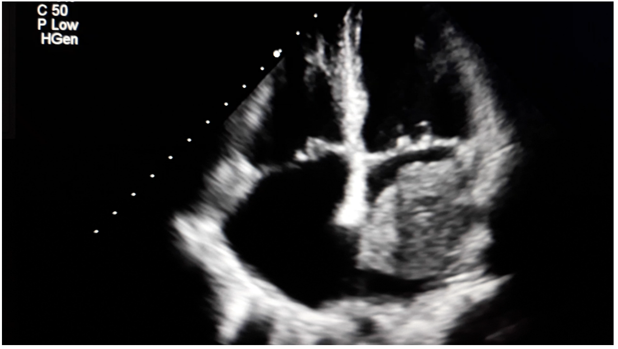
Introduction. Myxomas are the most common cavity primary tumor of the heart. Cardiac myxoma is 50% benign heart tumors [1,2]. In most cases, 75-80% cases, myxoma grows in the left atrium at the mitral ring or in the area of the atrium at the foramen ovale, 20% in the right atrium and in 5-8% in the ventricules. [3,4]. Myxoma usually grows on a so-called stem (a narrowed part of the tumor at the base) or attached to a wide base. This article presents a clinical case of a patient who was diagnosed with left atrial myxoma on echocardiography. Echocardiography is the most commonly used method in diagnostics. Accuracy 95% in the diagnosis of myxoma [9]. Transesophageal echocardiography (TEE) is 100% accurate study in the diagnosis of myxoma [1]. After a diagnosis of myxoma by imaging, surgical removal of the tumor should be performed as soon as possible. The results of surgical treatment are usually very good.
Aim: to present the clinical case in the Kaunas hospital of the Lithuanian University of Health Sciences. To select and analyze the causes, diagnostic and treatment recommendations of left atrial myxoma provided by experts.
Methods: the review of literature was conducted using medical databases selecting publications investigating left atrial myxoma.
Results: after the analysis of the literature, the main causes, diagnostics and treatment methods of left atrial myxoma are presented.
Conclusions: Myxomas are the most common cavernous benign heart tumor. Usually in 75-80% of cases myxoma grows in the left atrium. The clinical diagnosis of myxoma should be considered in all patients during clinical monitoring for symptoms of dyspnea, especially onset of sleep, nocturnal dyspnea, pulmonary edema, cough, hemoptysis and general fatigue.
Keywords: atrial myxoma, clinical case, tumor.