Rasita Pavilionė1, Laura Dobrovaitė2, Gabija Bagužytė2, Paulina Tekoriutė2
1Republic Hospital of Klaipeda, Klaipeda, Lithuania,
2Lithuanian University of Health Sciences, Academy of Medicine, Faculty of Medicine, Kaunas, Lithuania
Abstract
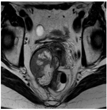
Endometriosis is a benign gynecological disease that affects 10-15% of reproductive age women. Endometriosis is defined as the presence of endometrial-like tissue outside the uterine cavity and can lead to infertility, dysmenorrhea, and chronic pain. Endometriosis involves bowels in about 8-12% of cases and in 90% it affects colon and rectum. Endometriotic lesions of bowel are associated with cyclic bowel alterations, dyschezia, rectal bleeding and obstipation. The most commonly used imaging techniques to identify and characterize endometriosis lesions are transvaginal sonography and magnetic resonance imaging. Aim: To present a clinical case of rectal endometriosis diagnosed in Republic Hospital of Klaipeda and review the latest scientific literature of this topic. Methods: A review of the literature using “PubMed” database was performed. Publications, researching the problem of endometriosis, were selected and a clinical case of rectal endometriosis was presented. Conclusions: Endometriosis is a challenging condition affecting quality of life in young women. This disease can affect a variety of organs which leads to non-specific clinical manifestation. Due to these doctors of different fields, including radiology, obstectrics and gynecology, gastroenterology, urology, family medicine and etc., can face difficulties in diagnosing this disease. In this article we present a clinical case of rectal endometriosis with imaging characteristics similar to rectal cancer.
Keywords: endometriosis, bowel endometriosis, transvaginal ultrasonography, magnetic resonance imaging.